Schedule Appointment
ADVANCED TECHNOLOGY
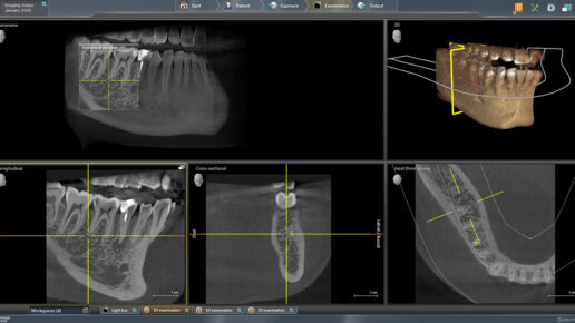
Axeos 3D/2D digital X-Ray
Dr. Francis recently incorporated the Axeos 3D/2D digital X-Ray machine and its software is the latest addition to our practice.
Axeos uses intelligent low-dose exposure to capture high-quality images while providing easy-to-use features to enhance patient comfort, such as smart height adjustment and quick scan times, that lead to exceptional patient experiences with high inflection prevention standards.
Adjustable ambient lighting helps to create a more pleasant, calming atmosphere for our patients. This technology integrates seamlessly with our other software and imaging products to create a truly digital workflow. This gives us more control for better outcomes and patient comfort for many procedures including implants, crowns, veneers and bridges.
We want our patients to receive the best dental care possible, that’s why we invested in the latest technology in dental diagnostics, the New Axeos 3D X-Ray.
DIGITAL IMAGING
Dr. Francis chooses carefully which and when radiographs are taken. There are many guidelines that we follow. Radiographs allow us to see everything we cannot see with our own eyes. Radiographs enable us to detect cavities in between your teeth, determine bone level, and analyze the health of your bone. We can also examine the roots and nerves of teeth, diagnose lesions such as cysts or tumors, as well as assess damage when trauma occurs.
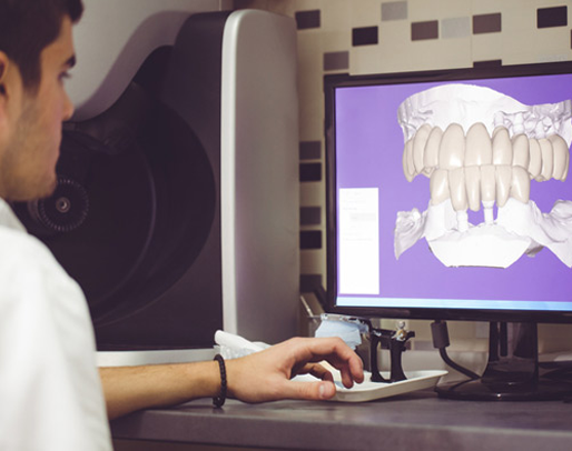
Dental radiographs are invaluable aids in diagnosing, treating, and maintaining dental health. Exposure time for dental radiographs is extremely minimal. Dr. Francis utilizes Digital Imaging Technologies within the office. With digital imaging, exposure time is about 50 percent less than compared to traditional radiographs. Digital imaging can also help us retrieve valuable diagnostic information. We may be able to see cavities better.
Digital imaging allows us to store patient images and enables us to quickly and easily transfer them to specialists or insurance companies.
LASER DENTISTRY
For patients who do not look forward to needles, drilling, or numbness, Laser Dentistry may be the right choice.
Laser dentistry is one of dentistry’s latest advances. The laser delivers energy in the form of light. Depending on the intended result, this energy travels at different wavelengths and is absorbed by a “target.” In dentistry, these targets can be enamel, decay, gum tissue, or whitening enhancers. Each one absorbs a different wavelength of light while reflecting others. Laser dentistry can be used for both tooth and soft tissue-related procedures. Oftentimes no local anesthesia is required. Unlike the dental drill, with laser dentistry there is no heat or vibration, making the procedure quite comfortable for most patients. For soft tissue (surgical) procedures it eliminates the need for suturing and healing is much faster.
Lasers can be used to diagnose cavities. They can find hidden decay in teeth in the early stages, and in some cases, the decay can be reversed through hygiene and fluoride treatment and may never need filling.
AREAS OF DENTAL CARE THAT BENEFIT FROM LASER TECHNOLOGY:
- Cavity diagnosis and removal
- Curing, or hardening, bonding materials
- Whitening teeth
- Periodontal, or gum related, care
- Pediatric procedures
- Apthous Ulcer treatment (canker sore)
- Frenectomy (tongue-tie release) without anesthesia or sutures
- Root canals and apicoectomies
- Crown lengthening, gingivectomy and other gum corrections
Dental lasers have been shown to be safe and effective for treating both children and adults.
We use the Gemini laser from Ultradent. The Gemini 810 + 980 diode laser is the first dual-wavelength soft tissue diode laser available in the United States. The unique dual-wavelength technology combines the optimal melanin absorption of an 810-nanometer wavelength diode laser with the optimal water absorption of a 980-nanometer wavelength diode laser.
INTRAORAL CAMERA
Many patients, especially younger patients, are very familiar with the latest technology and are comfortable with the high-tech practice. Computers and TV screens are their primary method of information processing.
Dr. Francis utilizes intraoral camera technology that helps enhance your understanding of your diagnosis. An intraoral camera is a very small camera – in some cases, just a few millimeters long. An intraoral camera allows our practice to view clear, precise images of your mouth, teeth and gums, in order for us to accurately make a diagnosis. With clear, defined, enlarged images, you see details that may be missed by standard mirror examinations. This can mean faster diagnosis with less chair-time for you!
Intraoral cameras also enable our practice to save your images in our office computer to provide a permanent record of treatments. These images can be printed for you, other specialists, and your lab or insurance companies.

